
Written by
Laura Elizabeth Lansdowne
Sponsored by
Cancer is a complex disease that stimulates both functional and compositional changes to the immune system. While our immune cells are able to detect and destroy cancer by recognizing neoantigens ‒ molecular features on the surface of cancer cells ‒ sometimes they seem to turn a blind eye and in some instances, tumors actually manipulate immunological processes to their advantage.
In this immersive article, we’ll take a tour through the tumor microenvironment, highlighting various immune cells along the way, and explore how components of the immune system can be exploited therapeutically to tackle the disease.
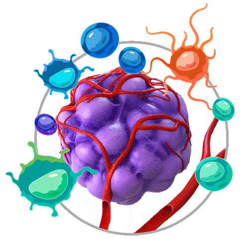
In addition to cancer cells, tumors comprise a variety of factors, including intricate networks of lymph and blood vessels, stromal cells, pericytes, extracellular matrix and components of the immune system ‒ giving rise to the tumor microenvironment (TME).
The immune response can be generally divided into the innate and adaptive immune systems, both contributing to the recognition and elimination of cancer cells.
While these immune cells may initially be recruited to the tumor site to play a host-protective role, their anticancer functions can be suppressed in response to tumor-derived signals and they may be coerced into a tumor-promoting role, encouraging tumor growth, invasion and metastasis.
Natural killer (NK) cells are typically found in the bone marrow and lymphatic tissues. NK cells mediate immunosurveillance of the tumor and migrate to the TME in response to secretion of chemokines by dendritic cells.
NK cells typically destroy cancer cells by releasing perforin and granzymes to induce apoptosis and can also influence adaptive antitumor immune responses by producing chemokines and cytokines.
On the flip side, soluble factors ‒ produced by different tumor-infiltrating immune cells and/or cancer cells ‒ may adversely affect NK-cell activity.
Cancer cells can also increase expression of MHC class I molecules on their surface to inhibit NK-cell activity and may decrease expression of certain ligands to avoid recognition by NKs.
B cells are typically found in the lymph nodes as well as in organized aggregates of immune cells that form in nonlymphoid tissues called tertiary lymphoid structures located close to the TME. B cells are known to play both a tumor-promoting and host-protective role. They can present tumor-associated antigens (TAAs) to T cells as well as produce antibodies against TAAs.
However, they are also able to inhibit T-cell responses via production of immunosuppressive cytokines (e.g., IL-10). B cells that express IgG4 and produce proangiogenic factors have also been shown to promote development of new blood vessels (angiogenesis), this in turn improves the tumor’s supply of oxygen and nutrients, and encourages proliferation and metastasis.
T-cell receptors trigger a cell-intrinsic signaling event upon association with a TAA or tumor-specific antigen (TSA). This causes the T cells to differentiate into cytotoxic effector cells that can destroy the cancer cells. However, when antigen-experienced T cells are repeatedly subjected to the same antigen or exposed to specific intrinsic factors, it can cause them to become dysfunctional.
Various T-cell populations exist within the TME and tumor margin. CD8+ T cells, CD4+ Th1 cells and γδ T cells are usually associated with a good prognosis, while FOXP3+ Treg cells, CD4+ Th2 cells and Th17 cells are associated with a poor prognosis.
Tumor-associated macrophages (TAMs) are present in most human cancers and are considered tumor-promoting. TAMs are able to produce proangiogenic factors and typically migrate to necrotic regions of the TME where oxygen levels are low, in response to hypoxia-induced chemoattractants (e.g., VEGF).
Dendritic cells (DCs) play a key role in antigen processing and presentation, and provide stimulation and soluble factors that shape T-cell activity. However, cancer cells can exploit DCs to promote immune tolerance. DCs are unable to trigger an immune response to TAAs in the absence of costimulatory signals, inducing T-cell anergy – the functional inactivation of T cells.
Dendritic cells (DCs) play a key role in antigen processing and presentation, and provide stimulation and soluble factors that shape T-cell activity. However, cancer cells can exploit DCs to promote immune tolerance. DCs are unable to trigger an immune response to TAAs in the absence of costimulatory signals, inducing T-cell anergy – the functional inactivation of T cells.
Myeloid-derived suppressor cells (MDSCs) are considered to be inhibitory immune cells, releasing large amounts of cytokines (e.g., IL-10). They also suppress host immune responses by releasing reactive oxygen species and nitric oxide. MDSCs can provoke macrophages to switch to a TAM-like phenotype, and are able to differentiate into TAMs themselves.
Myeloid-derived suppressor cells (MDSCs) are considered to be inhibitory immune cells, releasing large amounts of cytokines (e.g., IL-10). They also suppress host immune responses by releasing reactive oxygen species and nitric oxide. MDSCs can provoke macrophages to switch to a TAM-like phenotype, and are able to differentiate into TAMs themselves.
Many different immunotherapeutic strategies, or “immunotherapies”, have been designed to treat cancer. Their goal is to boost or restore the patient’s immune response to cancer cells, which may have been suppressed, so that they are able to eliminate the disease.
While immunotherapies have a common goal – to shift the balance back in favor of immune protection – the mechanisms they use to achieve this can be quite different. For example, they may work to increase the number of T cells, encourage antigen presentation, trigger activation of the innate immune system or modulate “immune checkpoints” – which are often manipulated by cancer cells to shield themselves from the immune system.
Some immunotherapies are preventive rather than therapeutic. Preventive immunotherapies aim to reduce the risk of disease, for example, by protecting a patient from infection with a cancer-causing virus or “oncovirus” (e.g., specific types of human papillomavirus).
Immune-modulating therapies influence the level of activity of the immune system, by stimulating or suppressing it. They can be broadly split into four types.
1. Checkpoint inhibitors block immune checkpoints
2. Cytokines allow immune cells to communicate effectively
3. Agonists promote adaptive immune responses
4. Adjuvants activate innate immune pathways
Adoptive cell therapies, or cellular immunotherapies, use immune cells to treat cancer. There are two main approaches, immune cells are either isolated, expanded and reintroduced into the patient or genetically modified to enhance their ability to kill cancer cells and then reintroduced into the patient.
Adoptive cell therapies include:
1. Tumor-infiltrating lymphocyte (TIL) therapy
2. Engineered T cell receptor (TCR) therapy
3. Chimeric antigen receptor (CAR) T-cell therapy
4. Natural killer (NK) cell therapy
Antibodies are produced by B cells in response to exposure to a specific antigen. Antibodies can be developed to target antigens that are typically found in greater numbers on the surface of cancer cells. Antibody therapies are classed as “passive” if they target cancer cells directly without involving immune cells or “active” if they require assistance from the immune system.
Oncolytic viruses are modified versions of viruses that are designed to target and destroy cancer cells and can also induce systematic antitumor immune responses. These viruses can be engineered to reduce their ability to infect healthy cells and they can also be used as carriers to transport therapeutic payloads to diseased cells. All oncolytic virus therapies can be administered via intratumoral injection, but some can also be delivered systemically (e.g., Myxoma virus).
Cancer vaccines can be categorized into three main types:
1. Preventive ‒ alert the immune system to the presence of an oncovirus so that it can clear the virus before it is able to cause an infection
2. Therapeutic ‒ strengthen a patient's immune responses, induce tumor regression and kill cancer cells, and establish lasting antitumor memory
3. Personalized ‒ target unique antigens (neoantigens) on the surface of cancer cells that result from specific mutations. The immune response is directed exclusively at cancer cells, reducing potential adverse effects.
Therapeutic monoclonal antibodies are one of the fastest-growing classes of drugs, targeting many disease areas including cancer, autoimmune disorders, chronic inflammation and infectious disease.
Traditional antibody screening workflows can be labor-intensive, may not support high-throughput applications, don’t allow for direct antibody comparison and need large amounts of reagents.
Download this whitepaper to discover how high-throughput flow cytometry can provide quantitative data on: